Drawing Of The Uterus
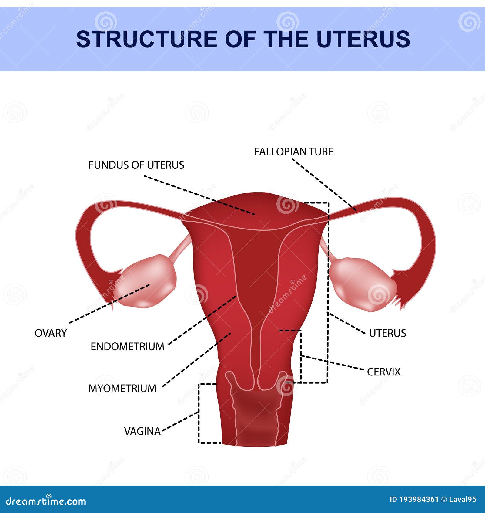
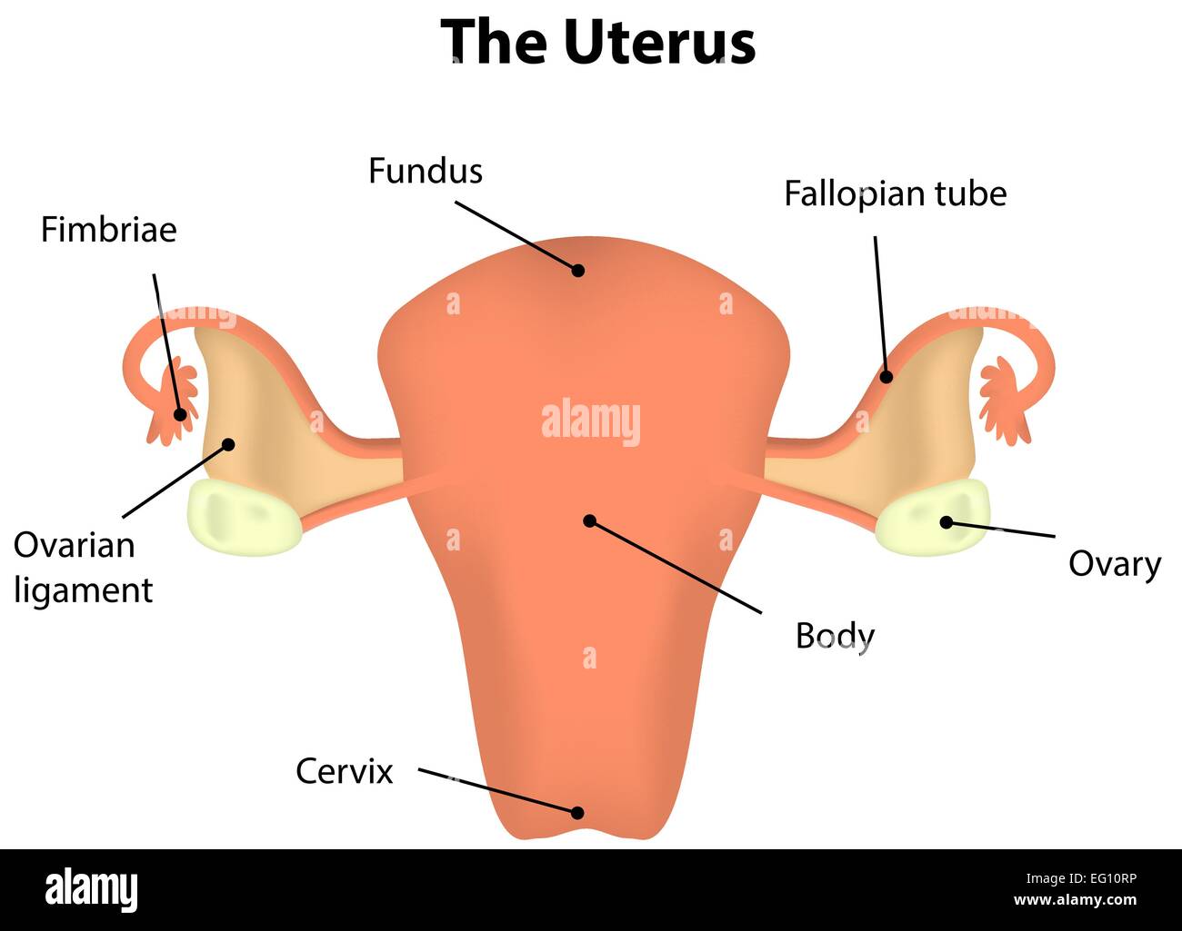
Drawing Of The Uterus - It's also called the womb. A smaller sketch of the same, and of details of the placenta and uterus; The uterus has three layers of muscle and is one of the strongest muscles in the body. The female gonads, the ovaries produce ova. Protein concentrations of each homogenate were determined by lowry assay. Drawing shows the uterus, myometrium (muscular outer layer of the uterus), endometrium (inner lining of the uterus), ovaries, fallopian tubes, cervix, and vagina. However, about 4% of females have a uterus that has a different shape. The width of the organ varies; Certain conditions and diseases of the uterus can cause painful symptoms that require medical treatment. This organ holds and nourishes a developing fetus, if an egg was properly fertilized. It’s hollow and muscular and sits between your rectum and bladder in your pelvis. Your corpus is the larger part of your uterus that expands during pregnancy. While its anatomy sounds simple, its histology is more complicated. The cervix and the corpus. It is generally about 6 cm wide at the fundus and only half this distance at the isthmus. Protein concentrations of each homogenate were determined by lowry assay. It is about the size of a fist. The uterus is where a fetus develops during pregnancy. The uterus has three layers of muscle and is one of the strongest muscles in the body. 101 labeled illustrations of the female genital system (ovaries, uterine tubes, uterus, vagina, vulva, clitoris) and pelvic cavity (bladder, rectum, pelvic diaphragm,. Web anatomy of the female reproductive system; The uterus has three layers of muscle and is one of the strongest muscles in the body. Web the uterus is 6 to 8 cm (2.4 to 3.1 inches) long; It's also called the womb. Certain conditions and diseases of the uterus can cause painful symptoms that require medical treatment. It is generally about 6 cm wide at the fundus and only half this distance at the isthmus. The cervix and the corpus. Web your uterus is divided into two parts: Web the uterus is a small, muscular organ in females that stretches to accommodate a growing fetus during pregnancy. A drawing of a fetus in utero, with the uterus. A drawing of a fetus in utero, with the uterus opened out; A diagram demonstrating binocular vision; Its wall thickness is approximately 2 to 3 cm (0.8 to 1.2 inches). Web the uterus is a female secondary sex organ located within the pelvis. It's in your lower belly (pelvic area). Web the uterus is a female secondary sex organ located within the pelvis. It's in your lower belly (pelvic area). It is part of the female reproductive system. Web anatomy of the female reproductive system; This organ holds and nourishes a developing fetus, if an egg was properly fertilized. Web the uterus is approximately the shape and size of a pear and sits in an inverted position within the pelvic cavity of the torso. It is usually present in. This organ holds and nourishes a developing fetus, if an egg was properly fertilized. The female reproductive organs include several key structures, such as the ovaries, uterus, vagina, and vulva.. It's in your lower belly (pelvic area). Web the uterus is a female secondary sex organ located within the pelvis. When one matures, it is released down into a. It's also called the womb. It is connected distally to the vagina, and laterally to the uterine tubes. Web the presence of regular uterine contractions (>3 contractions in a 10 minute period) with advanced cervical dilation (>3 centimeters.) tissue samples were frozen upon removal and remained so until homoginized on ice. Your uterus is connected to the fallopian tubes. The uterus is located in the lower belly area between the hips (pelvis), through the vagina just past the. It's sometimes called the womb. Web the uterus is an organ in the lower belly (abdomen) or pelvis. It is located along the body's midline posterior to the urinary bladder and anterior to the rectum. A drawing of a fetus in utero, with the uterus opened out; Anatomy atlas of the female pelvis: Carry eggs from the ovaries to the uterus. It is about the size of a fist. It's where a baby grows. It is located posterior to the urinary bladder and is connected via the cervix to the vagina on its inferior border and the fallopian tubes along its superior end. When one matures, it is released down into a. Web the uterus is approximately the shape and size of a pear and sits in an inverted position within the pelvic cavity of the torso. Notes on embryology, on relief in painting and on mechanics. The female reproductive organs include several key structures, such as the ovaries, uterus, vagina, and vulva. Web your uterus is divided into two parts: The. It consists of several anatomical parts, such as the cervix, isthmus, and body. Web the uterus is a female secondary sex organ located within the pelvis. It's also called the womb. Web the anatomy of the uterus consists of the following 3 tissue layers (see the following image): The uterus is where a fetus develops during pregnancy. The cervix and the corpus. Your corpus is the larger part of your uterus that expands during pregnancy. While its anatomy sounds simple, its histology is more complicated. Web your uterus is divided into two parts: It's in your lower belly (pelvic area). The female gonads, the ovaries produce ova. Web the uterus, also known as the womb, is an about 8 cm long hollow muscular organ in the female pelvis and lies dorsocranially on the bladder. It is located posterior to the urinary bladder and is connected via the cervix to the vagina on its inferior border and the fallopian tubes along its superior end. However, the proteome of prp in mares, particularly those susceptible to pbie, remains. A diagram demonstrating binocular vision; Notes on embryology, on relief in painting and on mechanics.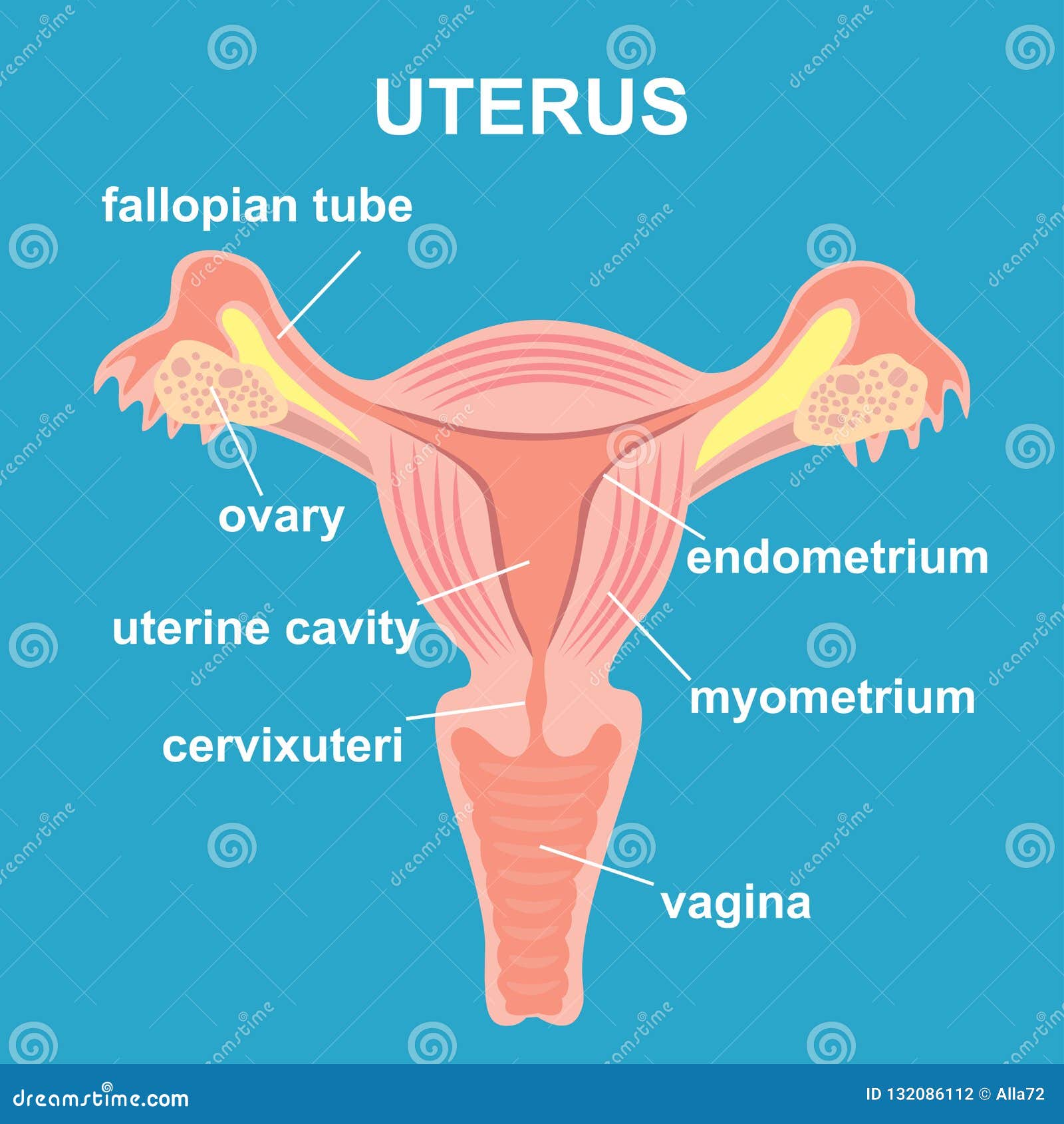
Uterus and Ovaries, Organs of Female Reproductive System Stock Vector
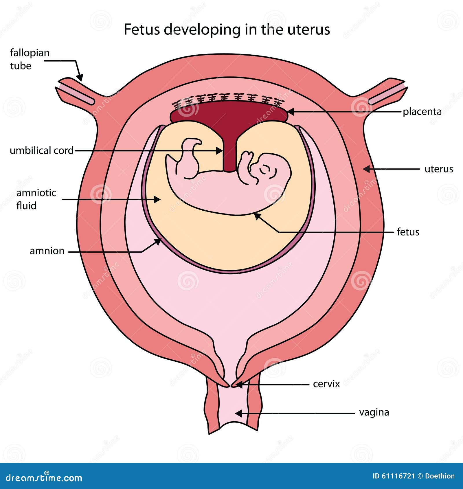
Diagram Of Uterus With Labels
![]()
Vector Isolated Illustration of Uterus Stock Vector Illustration of
Anatomy Of Ovarian Cycle RoyaltyFree Illustration
Uterus Labeled Diagram Stock Vector Image & Art Alamy
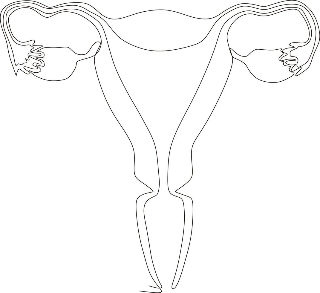
continuous line art drawing of female reproductive uterus 16818780

Contour Anatomical Sketch of the Uterus. Healthy Female Body. Woman

dibujo de arte de línea continua del útero reproductivo femenino

The Uterus Structure Location Vasculature TeachMeAnatomy
![]()
Premium Vector Anatomical human uterus vector line icon. hand drawn
It Is About The Size Of A Fist.
Its Wall Thickness Is Approximately 2 To 3 Cm (0.8 To 1.2 Inches).
In The Human, The Lower End Of The Uterus Is A Narrow Part Known As The Isthmus That Connects To The Cervix, Leading To The.
Web 240 Uterus Drawing Stock Illustrations And Clipart.
Related Post: